
 RadCases Head and Neck Imaging
by
RadCases Head and Neck Imaging
by
 Head and Neck Imaging
by
Head and Neck Imaging
by
The head and neck region is one of the most complex anatomical areas, housing critical structures for communication, sensation, respiration, and circulation. This section of the guide provides curated resources to help you explore the bones, muscles, nerves, glands, and vascular systems of the head and neck through multiple learning modalities.
Whether you're studying for a clinical exam, preparing a lesson plan, or simply curious about human anatomy, you'll find tools here to support your learning style—visual, text-based, or interactive.
Begin your exploration with foundational resources that introduce the major anatomical structures of the head and neck. These include the skull, facial muscles, cranial nerves, salivary glands, and lymphatic drainage pathways.
 Gray's Anatomy for Students
by
Concise, readable text and an outstanding art program make Gray's Anatomy for Students, 5th Edition, your go-to text for essential information in human anatomy. This fully revised volume focuses on the core information medical students need to know, in an easy-access format and with additional multimedia content to facilitate effective study and mastery of the material. A team of expert authors share a wealth of diverse teaching and clinical experience-all enhanced by more than 1,000 innovative, original illustrations by renowned illustrators Richard Tibbitts and Paul Richardson, who capture anatomical features with unrivalled clarity. Helps you understand the practical applications of anatomical concepts through unique coverage of surface anatomy, correlative diagnostic images, and clinical case studies. Contains increased representation of diverse population groups throughout, incorporating a wider range of skin tones and important clinical considerations related to transgender and intersex individuals. Presents anatomy logically by body region - as well as bonus updated eBook chapters for each major body system to facilitate learning from a different perspective. Includes new and improved online materials such as self-assessment questions, medical and physical therapy clinical cases, a unique Interactive Surface Anatomy tool, and more. Provides fully revised and updated clinical content including numerous new In the Clinic boxes, images, and correlates throughout that reflect the latest advances seen in clinical practice. New and updated Clinical Cases are included in the accompanying enhanced eBook. Features an updated neuroanatomy eBook chapter, so you can learn key aspects of this challenging topic in the context of general anatomy. Improves comprehension of complex cranial nerves with a visual map summarizing cranial nerve distribution and function. Offers schematic drawings for key structures and topics in every chapter, providing an additional, simplified approach to introduce each topic-ideal for quick initial understanding and as a guide for your own anatomy drawings. Enables you to quickly review the basic concepts from each chapter with Conceptual Overviews. An eBook version is included with purchase. The eBook allows you to access all of the text, figures and references, with the ability to search, customize your content, make notes and highlights, and have content read aloud. Evolve Instructor site with a downloadable image bank is available to instructors through their Elsevier sales rep or via request at https://evolve.elsevier.com.
Gray's Anatomy for Students
by
Concise, readable text and an outstanding art program make Gray's Anatomy for Students, 5th Edition, your go-to text for essential information in human anatomy. This fully revised volume focuses on the core information medical students need to know, in an easy-access format and with additional multimedia content to facilitate effective study and mastery of the material. A team of expert authors share a wealth of diverse teaching and clinical experience-all enhanced by more than 1,000 innovative, original illustrations by renowned illustrators Richard Tibbitts and Paul Richardson, who capture anatomical features with unrivalled clarity. Helps you understand the practical applications of anatomical concepts through unique coverage of surface anatomy, correlative diagnostic images, and clinical case studies. Contains increased representation of diverse population groups throughout, incorporating a wider range of skin tones and important clinical considerations related to transgender and intersex individuals. Presents anatomy logically by body region - as well as bonus updated eBook chapters for each major body system to facilitate learning from a different perspective. Includes new and improved online materials such as self-assessment questions, medical and physical therapy clinical cases, a unique Interactive Surface Anatomy tool, and more. Provides fully revised and updated clinical content including numerous new In the Clinic boxes, images, and correlates throughout that reflect the latest advances seen in clinical practice. New and updated Clinical Cases are included in the accompanying enhanced eBook. Features an updated neuroanatomy eBook chapter, so you can learn key aspects of this challenging topic in the context of general anatomy. Improves comprehension of complex cranial nerves with a visual map summarizing cranial nerve distribution and function. Offers schematic drawings for key structures and topics in every chapter, providing an additional, simplified approach to introduce each topic-ideal for quick initial understanding and as a guide for your own anatomy drawings. Enables you to quickly review the basic concepts from each chapter with Conceptual Overviews. An eBook version is included with purchase. The eBook allows you to access all of the text, figures and references, with the ability to search, customize your content, make notes and highlights, and have content read aloud. Evolve Instructor site with a downloadable image bank is available to instructors through their Elsevier sales rep or via request at https://evolve.elsevier.com.
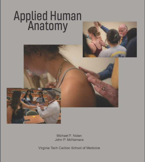 Applied Human Anatomy
by
Applied Human Anatomy
by
Visual learners grasp information best through images, diagrams, videos, and spatial representations. If you find yourself sketching structures or using color-coded notes, this section is for you.
You’ll benefit from:
RadCases Head and Neck Imaging
by
All the key Radiology cases for your rounds, rotations, and exams Head and Neck Imaging presents the challenging cases that are most likely to be encountered by residents and radiologists focusing on imaging of the head and neck. It helps radiologists correctly interpret images and thus quickly make initial diagnoses on both common and less common disorders of the head and neck. This book is also an effective review guide for those studying for the radiology board exams. Features of Head and Neck Imaging: Content covers the imaging of the temporal bone, skull base, orbits, paranasal sinuses, and all the structures of the head and neck Examples of critical cases that must be accurately diagnosed in daily practice and on exams Clearly labeled, high-quality images help you quickly absorb key findings RadCases contains cases selected to simulate everything that you'll see on your rounds, rotations, and exams. RadCases also helps you identify the correct differential diagnosis for each case--including the most critical. RadCases covers: Cardiac Imaging · Interventional Radiology · Musculoskeletal Radiology · Neuro Imaging · Thoracic Imaging · Pediatric Imaging · Gastrointestinal Imaging · Breast Imaging · Emergency Radiology · Nuclear Medicine · Ultrasound Imaging · Head and Neck Imaging · Genitourinary Imaging Each RadCases title features 100 carefully selected, must-know cases documented with clear, high-quality radiographs. The organization provides maximum ease of use for self-assessment. Each case begins with the clinical presentation on the right-hand page; simply turn the page for imaging findings, differential diagnoses, the definitive diagnosis, essential facts, and more. This RadCases book comes with a code providing access to additional online cases: 100 from this book plus 150 more cases. Learn your cases, diagnose with confidence, and pass your exams. RadCases. This print book includes complimentary access to a digital copy on https://medone.thieme.com. Publisher's Note: Products purchased from Third Party sellers are not guaranteed by the publisher for quality, authenticity, or access to any online entitlements included with the product.
Also known as verbal or linguistic learners, you thrive on reading and writing. You prefer detailed explanations, structured outlines, and written definitions.
You’ll benefit from:
Head and Neck Imaging
by
This book provides a practically applicable guide to the all the different imaging modalities used in the diagnosis and management of ENT & Head and Neck patients. It bridges the gap in understanding between surgeons treating ENT & Head and Neck conditions and radiologists who oversee the process of scan requests, interpretation and delivering reports that best inform the subsequent management. Chapters cover a variety of sub-specialist areas including plain films, ultrasound, computed tomography (CT), magnetic resonance imaging (MRI), auditory implantation, paediatrics, head and neck cancer, trauma, three dimensional (3D) reconstruction and rehabilitation including swallow. This book facilitates surgeons and radiologists to further develop their understanding of each other's perspectives on clinical decision-making and appropriately interpreting the outputs from a range of imaging modalities. Head and Neck Imaging: A Multi-Disciplinary Team Approachis a resource well-suited to all trainees, residents, consultants who use these techniques to treat patients with head and neck symptoms. Furthermore, it is vital for those individuals preparing for exams in disciplines such as ear nose and throat, maxillofacial surgery and radiology.
A core resource for health sciences programs, this database provides full-text access to authoritative biomedical journals indexed by the U.S. National Library of Medicine. Topics include clinical medicine, nursing, public health, and healthcare systems. Features Medical Subject Headings (MeSH) for precise searching, supporting evidence-based research, academic study, and professional practice.
You learn best by doing—through hands-on activities, simulations, and self-testing. If you enjoy quizzes, virtual labs, or building models, this section is for you.
You’ll benefit from:
An immersive, interactive learning tool that allows students to explore human anatomy through detailed 3D models and virtual dissections. Users can zoom, rotate, and dissect anatomical structures to study body systems in depth. Designed for college-level anatomy courses, the platform includes guided lessons, self-assessments, and integrated reference content from Gale. Ideal for both in-class demonstrations and independent study, it supports visual and kinesthetic learning styles.
Here are some engaging, hands-on activities to deepen your understanding of head and neck anatomy:
Compare Imaging Modalities: Use RadCases: Head and Neck Imaging to compare CT and MRI scans of the paranasal sinuses. Identify the same anatomical landmarks in both.
3D Dissection: Use Gale Interactive: Human Anatomy to rotate and dissect the skull. Focus on the cranial base and identify foramina and the structures that pass through them.
Create a Study Set: Use your favorite flashcard tool to build a set for head and neck arteries, including the branches of the external carotid artery.
Cross-Reference Anatomy and Imaging: Use Head and Neck Imaging: A Multi-Disciplinary Team Approach to match anatomical diagrams with clinical imaging. Try identifying the thyroid gland, lymph nodes, and salivary glands in different imaging planes.