
Explore the structure and function of the eye, including the eyelid, orbit, and lacrimal system. These resources provide foundational knowledge for students in health sciences, nursing, and biology.
 Anatomy of the Eyelid, Orbit, and Lacrimal System
by
Anatomy of the Eyelid, Orbit, and Lacrimal System
by
 Atlas of Orbital Imaging
by
Atlas of Orbital Imaging
by
 Anatomy of the eye and orbit : the clinical essentials
by
Anatomy of the eye and orbit : the clinical essentials
by
Visual learners thrive when information is presented in a way that they can see and interpret spatially. If you find that diagrams, illustrations, videos, and color-coded notes help you understand complex topics, this section is for you. The anatomy of the eye is highly visual—its intricate structures and spatial relationships are best appreciated through high-quality images and interactive models. Here, you’ll find resources that allow you to explore the eye in 3D, view labeled diagrams, and watch animations that bring ocular anatomy to life.
Gale Interactive: Human Anatomy
Explore 3D models of the eye and surrounding structures.
Films on Demand
Search for “eye anatomy” to find educational videos.
Netter Images
A comprehensive collection of medical illustrations.
An immersive, interactive learning tool that allows students to explore human anatomy through detailed 3D models and virtual dissections. Users can zoom, rotate, and dissect anatomical structures to study body systems in depth. Designed for college-level anatomy courses, the platform includes guided lessons, self-assessments, and integrated reference content from Gale. Ideal for both in-class demonstrations and independent study, it supports visual and kinesthetic learning styles.
A comprehensive streaming video platform offering over 45,000 full-length academic videos and more than 341,000 video clips across 25+ core subject areas. Curated from over 1,200 international producers, the collection supports a wide range of disciplines including history, science, business, engineering, health, arts, and social sciences. Features include public performance rights, closed captioning, interactive transcripts, and tools for creating playlists, embedding clips, and generating citations.
If you prefer reading detailed explanations and writing things down to reinforce your learning, you’re likely a text-based learner. This section is tailored for those who absorb information best through written content—whether it’s textbooks, articles, or structured outlines. The resources here include comprehensive print and digital texts that explain the anatomy and physiology of the eye in depth. These materials are ideal for note-taking, outlining, and building a strong conceptual foundation for exams or clinical practice.
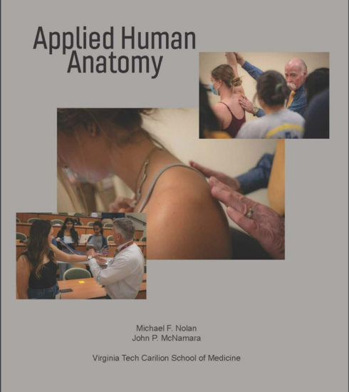 Applied Human Anatomy
by
Applied Human Anatomy
by
Provides access to a collection of over 125,000 academic e-books across a wide range of disciplines. Includes scholarly monographs, encyclopedias, dictionaries, and reference works. Content is curated and permanently owned by OhioLINK, with funding from member libraries and central support.
Interactive learners understand best by doing. If you enjoy hands-on activities, simulations, or testing your knowledge through quizzes and games, this section is designed for you. The eye is a dynamic organ, and exploring it through virtual dissections, 3D models, and interactive tools can make learning more engaging and memorable. These resources encourage active participation, helping you connect theory to practice through exploration and experimentation.
Put your knowledge into action with these interactive and practical exercises. These activities are designed to reinforce your understanding of ocular anatomy through exploration, comparison, and self-assessment.
Download a blank diagram of the eye from Get Body Smart or your textbook. Then:
Use the Atlas of Orbital Imaging (available through the Br. Edmond Drouin Library) to:
Log into Gale Interactive: Human Anatomy and:
Using content from Anatomy of the Eyelid, Orbit, and Lacrimal System:
Read a case study involving an eye condition (e.g., orbital cellulitis or glaucoma) from a textbook or database like CINAHL or MEDLINE. Then:
An immersive, interactive learning tool that allows students to explore human anatomy through detailed 3D models and virtual dissections. Users can zoom, rotate, and dissect anatomical structures to study body systems in depth. Designed for college-level anatomy courses, the platform includes guided lessons, self-assessments, and integrated reference content from Gale. Ideal for both in-class demonstrations and independent study, it supports visual and kinesthetic learning styles.
An essential database for nursing and allied health research, offering full-text access to a wide range of peer-reviewed journals, evidence-based care sheets, and continuing education materials. Ideal for nursing students, educators, and clinicians, it supports coursework, clinical decision-making, and scholarly research across disciplines such as public health, physical therapy, nutrition, and more.
A core resource for health sciences programs, this database provides full-text access to authoritative biomedical journals indexed by the U.S. National Library of Medicine. Topics include clinical medicine, nursing, public health, and healthcare systems. Features Medical Subject Headings (MeSH) for precise searching, supporting evidence-based research, academic study, and professional practice.